Intracellular integration of synthetic nanostructures with viable cells for controlled biochemical manipulation
Publications
Journal articles
This green picture has a life of its own. If you see it somewhere please let me know so I can add a link on this page to trace it.
Thank you
Email me at: amelech2@utk.edu
McKnight, T.E., A.V.
Melechko, G.D. Griffin, M.A. Guillorn, V.I. Merkulov, F.
Serna, D.K. Hensley, M.J. Doktycz, D.H. Lowndes, and M.L.
Simpson,
Nanotechnology, 2003. 14(5): p. 551-556
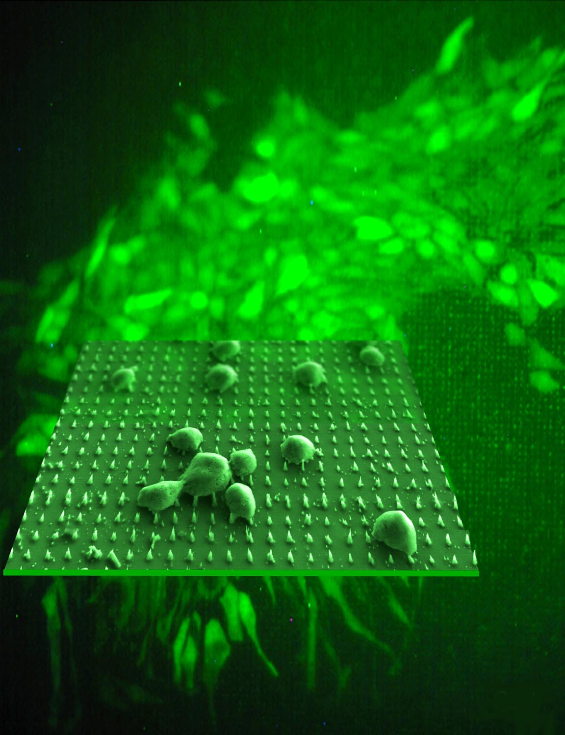
This image has been assembled in Adobe
Photoshop by overlaying an optical microscope photograph and scanning
electron micrograph that has been colorized and manipulated to display
perspective.
Front: Scanning electron micrograph of
chinese hamster ovary cells (CHO) following impalement on a nanofiber
array. Background: Optical microscope image of a transformed colony of
CHO expressing green fluorescent protein from nanofiber delivered
plasmids 22 days following impalement upon DNA modified nanofiber array.
Expecting Big Things from Nanostructures
NANOSTRUCTURES OFFER NEW APPROACH TO CELL MANIPULATION
Cancer Nanotech (National Institute of
Cancer) brochure
[local save]
National Institute of Biomedical Imaging and Bioengineering (found by Jason Heikenfeld)
www.nano.gov report on Nanobiotechnology (saved on this server)

The idea of impalefection is scribbled behind Tim McKnight's head on this photo